-
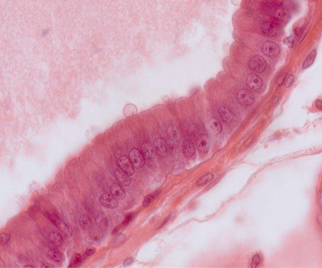
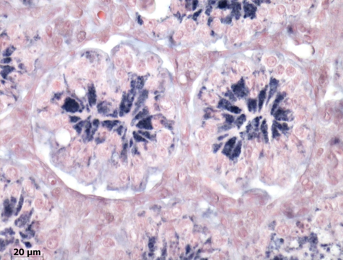
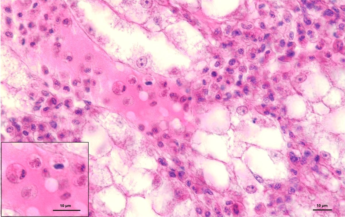
N. 1 - Cell Sunrise
Hematoxylin–eosin staining reveals the radiant morphology of prostasomes released by rat prostate cells. 100× magnification
-
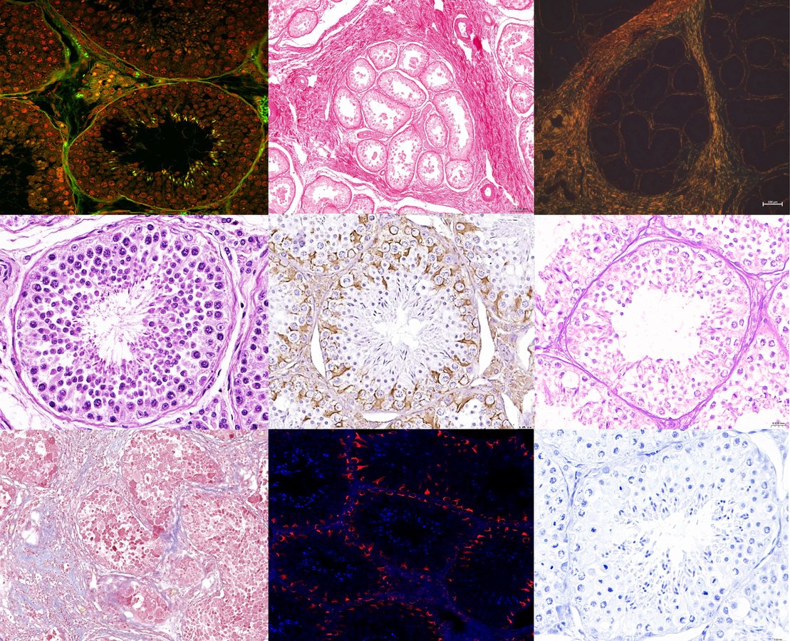
N. 2 - Pop Histology: omaggio a Marilyn
Composizione in chiave pop-art di immagini ottenute da sezioni istologiche di testicolo canino, ispirata all’estetica seriale di Andy Warhol. Da sinistra a destra, dall’alto in basso: Acridine Orange, Picrosirius Red in campo chiaro, Picrosirius Red in luce polarizzata, H&E, IHC vimentina, PAS, Tricromica di Masson, IF vimentina, Toluidine blue
-
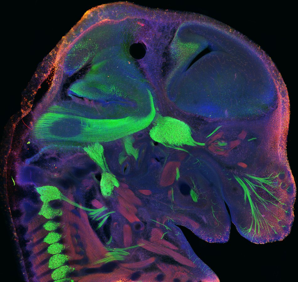
N. 3 - The glow of growing perception
Immagine al microscopio confocale di embrione murino allo stadio di E14.5 in cui sono visualizzati in verde i pathway sensoriali (dorsal root ganglia, gangli dei nervi trigemino, glossofaringeo e vago e nervi somatosensoriali dei whiskers) ed in rosso i fasci di muscolatura striata
-
N. 4 - Hey you, would you help me to carry the stone?
Saette di tirosina idrossilasi (TH) 🟡 solcano i neuroni dopaminergici della substantia nigra pars compacta di un modello murino trattato con rotenone. I bagliori del danno ossidativo 8-OH(d)G 🔵 illuminano la scena, mentre i nuclei ⚪ fluttuano silenziosi nel cuore di una tempesta neurobiologica.
-
N. 5 - Sertoli’s Secret Garden
In situ hybridization for the localization of VIP (Vasoactive intestinal peptide) mRNA in the testis of the lizard Podarcis siculus. Testis sections in the non-reproductive period were incubated with a DIG-labeled probes pecific for VIP mRNA. Positive hybridization signals appear as blue staining in the apical portion of Sertoli cells
-
N. 6 - Huston, we have a cell! - Cosmic trafficking inside Astro-Cell
Confocal microscopy image acquired using a ZEISS LSM 900 microscope equipped with a 63× objective. U251 glioblastoma cells were stained with DAPI (blue) and MitoTracker CMXRos (red) following 72 hour treatment with the autofluorescent f-N8 molecule (green)
-
N. 7 - Il giardino del nostro benessere / The Garden of Our Well-being
Villo duodenale con ghiandole trasversali, colorato con AB-PAS. L'immagine evidenzia l'organizzazione della mucosa intestinale e delle strutture ghiandolari, evocando un giardino microscopico in cui forma, colore e funzione contribuiscono al nostro benessere/ Duodenal villus with transverse glands, stained with AB-PAS. The image highlights the organization of the intestinal mucosa and glandular structures, evoking a microscopic garden where form, color and function contribute to our well-being
-
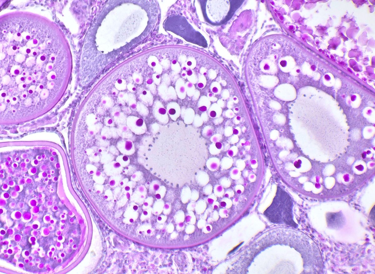
N. 8 - Pink reserves for a new life
Zebrafish cortical alveolar oocyte at magnification 10x stained with Periodic Acid Shiff which highlights vitellogenin granules, the gathering of resources that precedes the start of new life
-
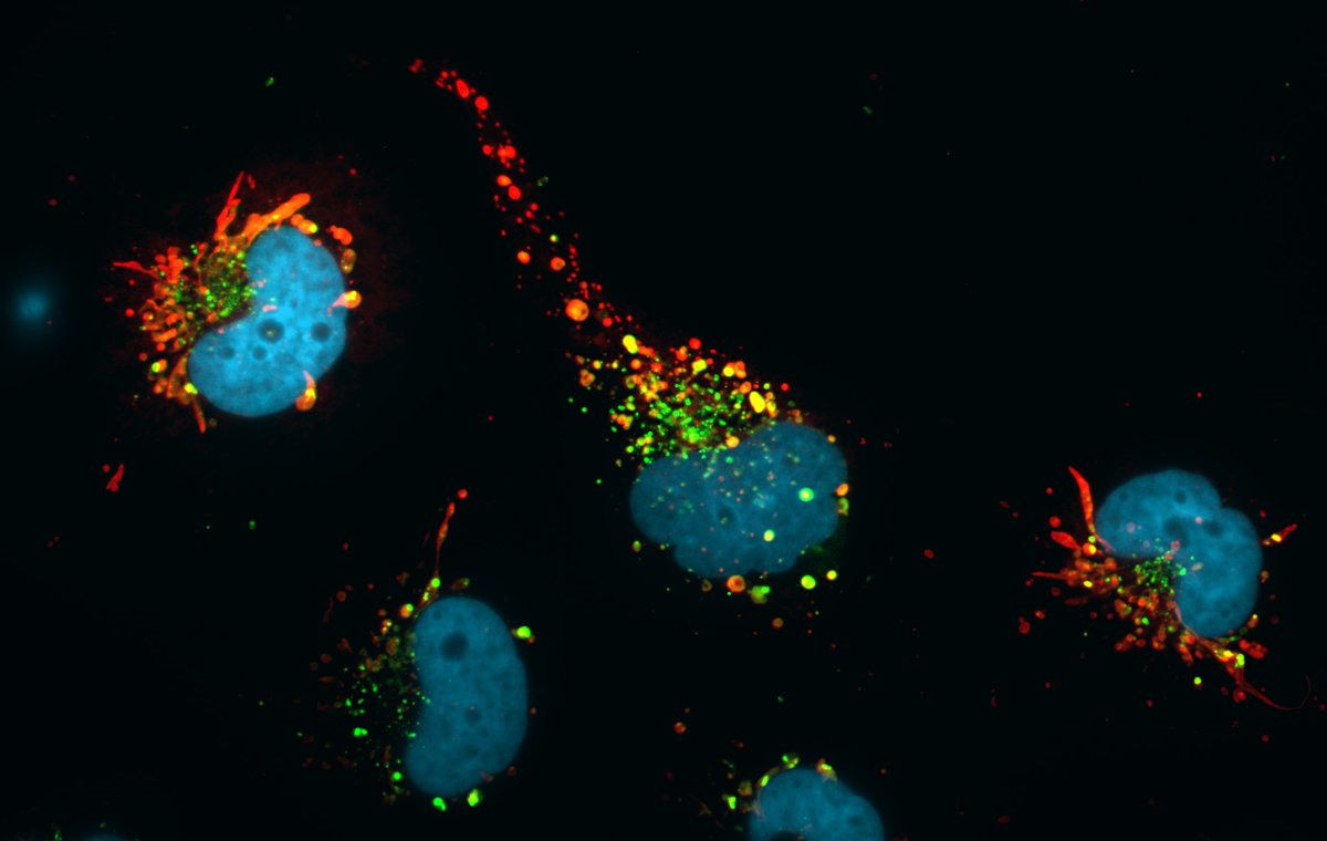
N. 9 - THunderstorm
When life gives you nanoplastics, pass them to a friend: immune cells caught in the act of transferring phagocytosed nanoplastic particles (red) through cytoplasmic protrusions, proving that even at the microscopic scale, nobody faces pollution alone. Cytoskeleton in green, nuclei in blue
-
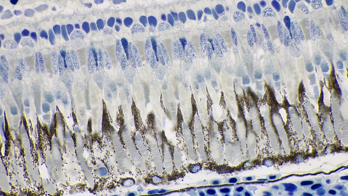
N. 10 - Neural Forest of Vision
Sezione istologica della retina osservata al microscopio ottico (100×) e colorata con blu di toluidina. L’organizzazione dei fotorecettori richiama l’immagine di una foresta, dove ogni “albero” contribuisce alla cattura della luce e alla costruzione della visione
-
N. 11 - Ragione e Sentimento
Sezione del telencefalo di Xenopus laevis colorata con blu di toluidina. Nelle pieghe del cervello anfibio si intrecciano le radici evolutive delle funzioni che, nell’uomo, associamo al pensiero e alle emozioni
-
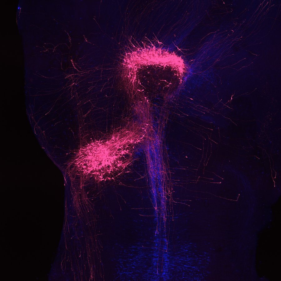
N. 12 - When Development Takes Flight
Espianto di neuroni serotoninergici provenienti da un embrione Tph2-GFP trapiantato su un host Ai14/Pet1-Cre. I neuroni e le fibre in crescita si espandono nel tessuto ospite disegnando spontaneamente la sagoma di un fenicottero, dove biologia dello sviluppo e immaginazione si incontrano
-
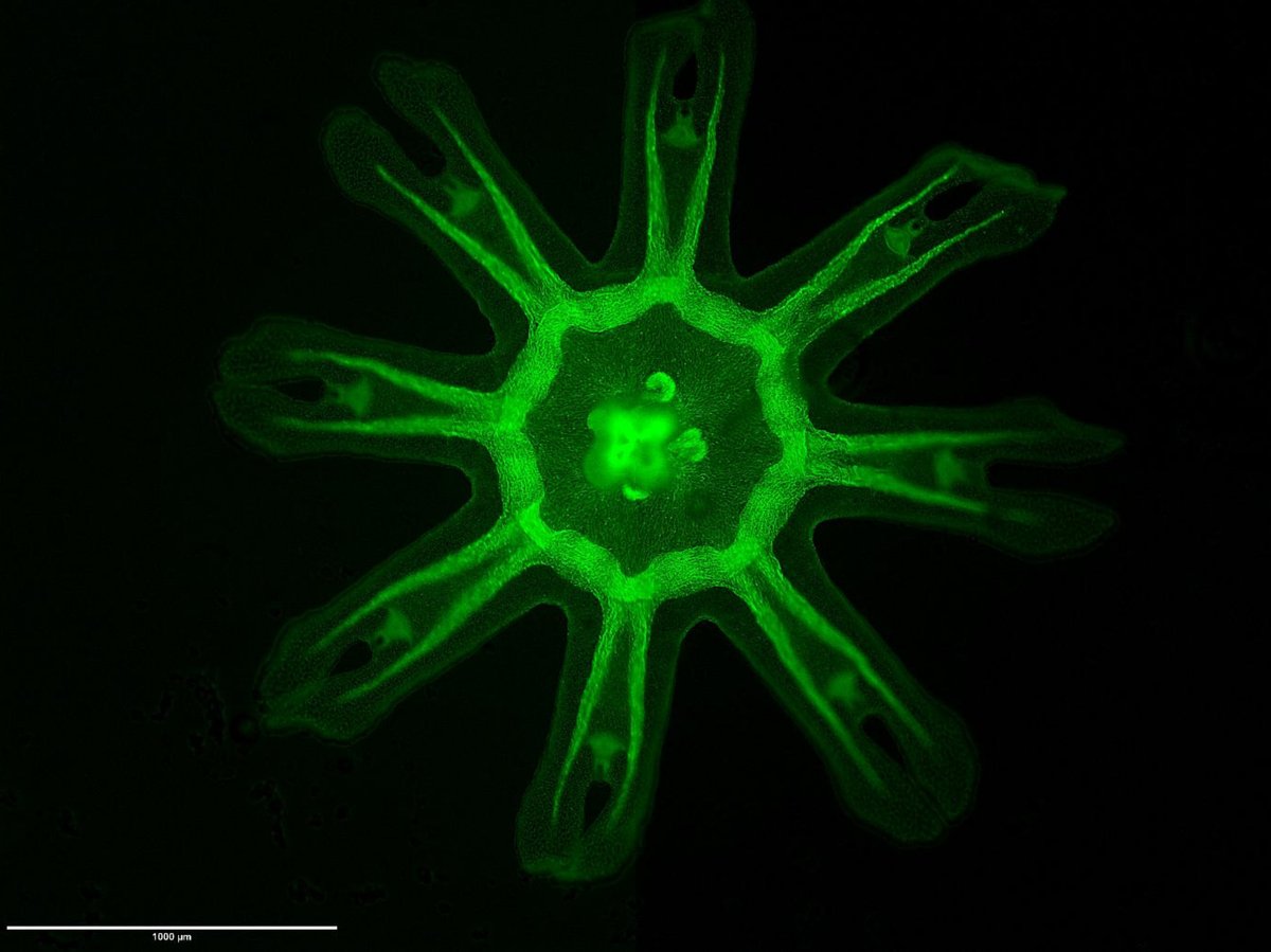
N. 13 - Four of Clubs!
A fluorescent image of an Aurelia aurita ephyra, lying flat with its eight bifid arms radiating outward toward the corners of the image and a luminous peduncle at the center. It depicts the juvenile stage just released from the polyp: eight arms, four pairs, foreshadowing the tetrameric symmetry of the adult jellyfish
-
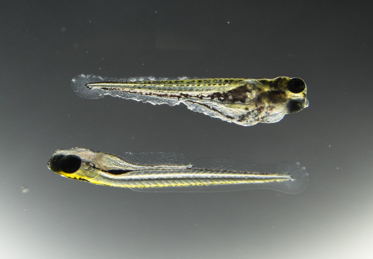
N. 14 - The unfortunate brother
Before they had any chance to experience the world, two zebrafish brothers were randomly assigned to different fates. This image reunites them: above, one bears the marks of a harsh chemical environment—oedema and incomplete fin development; below, his sibling developed normally under ideal conditions.
-
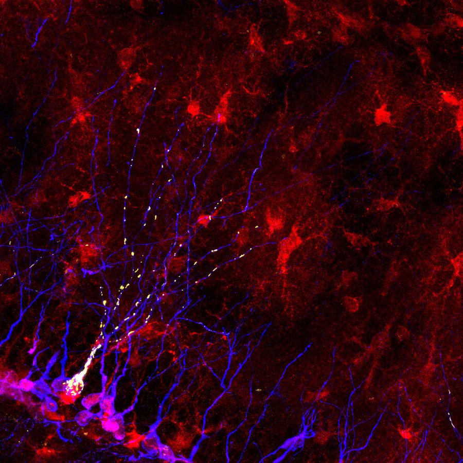
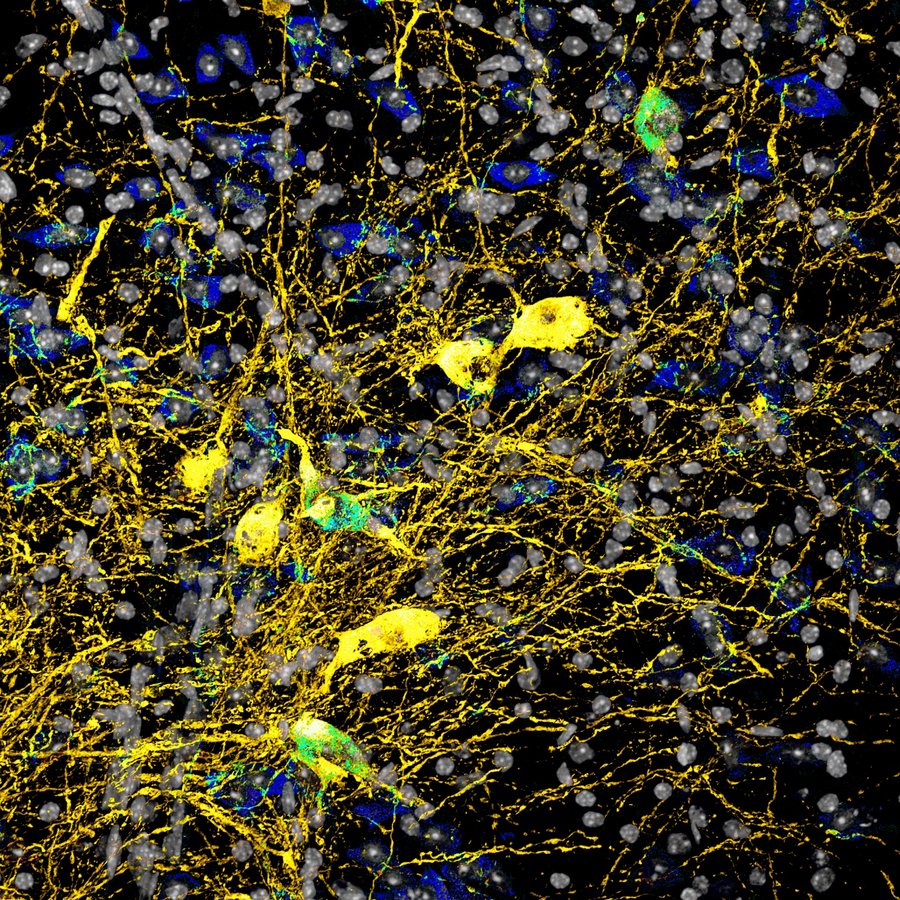
N. 15 - Costellazioni mitocondriali: l'energia che accende i nuovi neuroni
Immagine confocale della neurogenesi adulta ippocampale. Sullo sfondo rosso di astrociti e cellule staminali neurali (YFP+), si ramificano neuroni immaturi neogenerati (Doublecortina, blu). Un retrovirus codificante mitoDsRed svela in giallo la complessa architettura mitocondriale che "alimenta" lo sviluppo neuronale
-
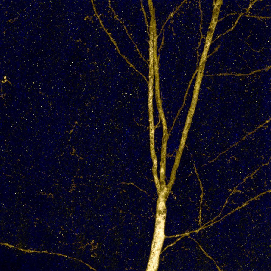
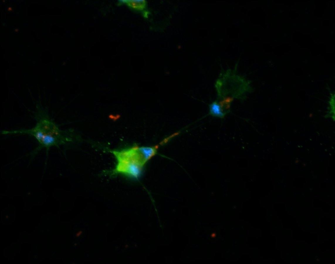
N. 16 - Nocturne for a Neuron
Dettaglio di albero dendritico di neurone piramidale marcato con GFP, acquisito mediante microscopia confocale ad alta risoluzione. I contatti sinaptici sono identificati tramite immunofluorescenza per PSD95, in blu e Bassoon, in giallo, rispettivamente marker della densità postsinaptica e della zona attiva presinaptica
-
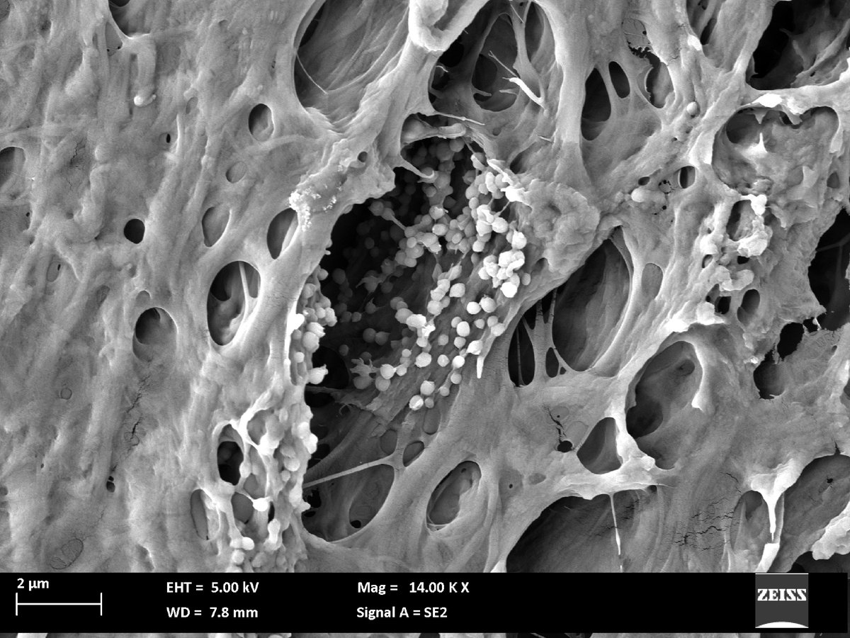
N. 17 - Let It Grow: The Secret Life of Vesicles
Scanning electron micrograph (SEM) showing clustered extracellular vesicles (EVs) nestled within the porous cavities of a nanostructured scaffold injected into a leech model. This high-resolution image captures the intricate vesicle-scaffold interface, highlighting their potential role in advancing regenerative medicine. Scale bar: 2 µm.
-
N. 18 - Hemocyte traffic is getting messy
In the kidney of a juvenile Pomacea canaliculata snail, different hemocyte morphotypes flow through wide hemolymph sinuses like smooth traffic on open roads. The pathway narrows between epithelial crypts, forcing the hemocytes to jam into crowded bottlenecks. There, they accumulate into apparently homogeneous "islets", i.e., hemocyte-resident sites in adult snails
-
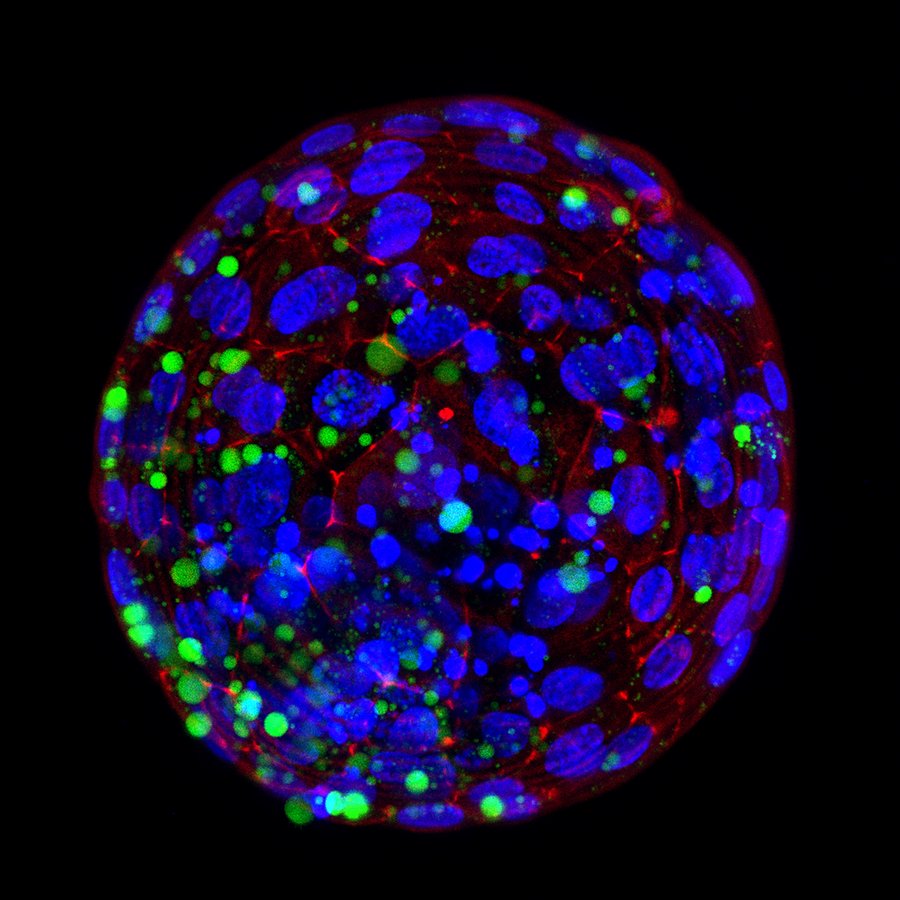
N. 19 - Bubble Blast
Immagine confocale di blastocisti bovina marcata in fluorescenza. Il citoscheletro cellulare appare in rosso (falloidina), i nuclei in blu (Hoechst) e le droplets lipidiche in verde (BODIPY). La struttura tridimensionale rivela l’architettura cellulare e la distribuzione lipidica durante le prime fasi dello sviluppo embrionale
-
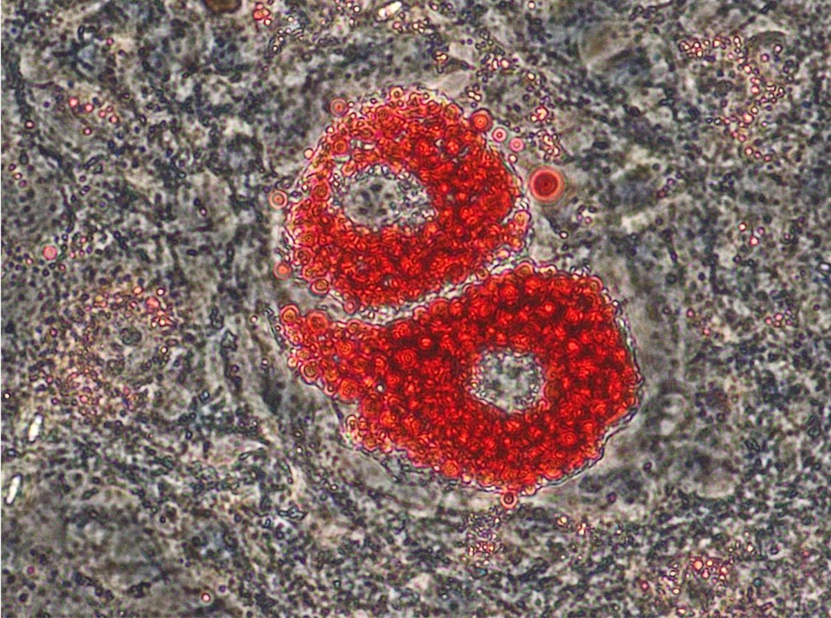
N. 20 - Cellular Couture: Adipocytes in Valentino Red
Oil Red O staining reveals lipid accumulation in two differentiating 3T3-L1 adipocytes, capturing a pivotal moment in adipogenesis. Image acquired using a 40x objective
-
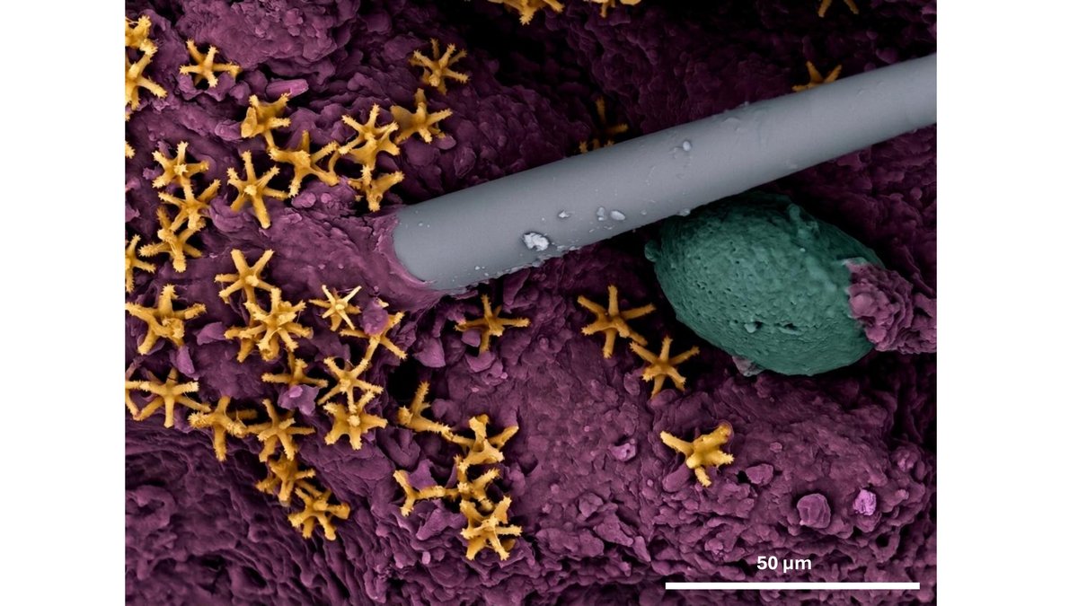
N. 21 - The sponge cosmos: oocyte in a constellation of spicules
The scanning electron microscopy (SEM) image shows the parenchyma (purple) of the sponge species Thethya meloni, with a clearly visible oocyte (green). Numerous micrasters (yellow), star-shaped skeletal elements, are distributed throughtout the matrix
-
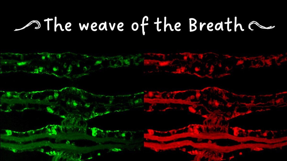
N. 22 - The weave of the Breath
A network of fluorescent signals spans the Mytilus galloprovincialis gills in response to treatment with persistent micropollutants like pharmacologically active compounds. Immunofluorescent labeling for serotonin (5-HT, green) and its receptor (5-HT3 R, red) reveals a hidden web of biological signals underlying the continuous and vulnerable dialogue between biota and environment
-
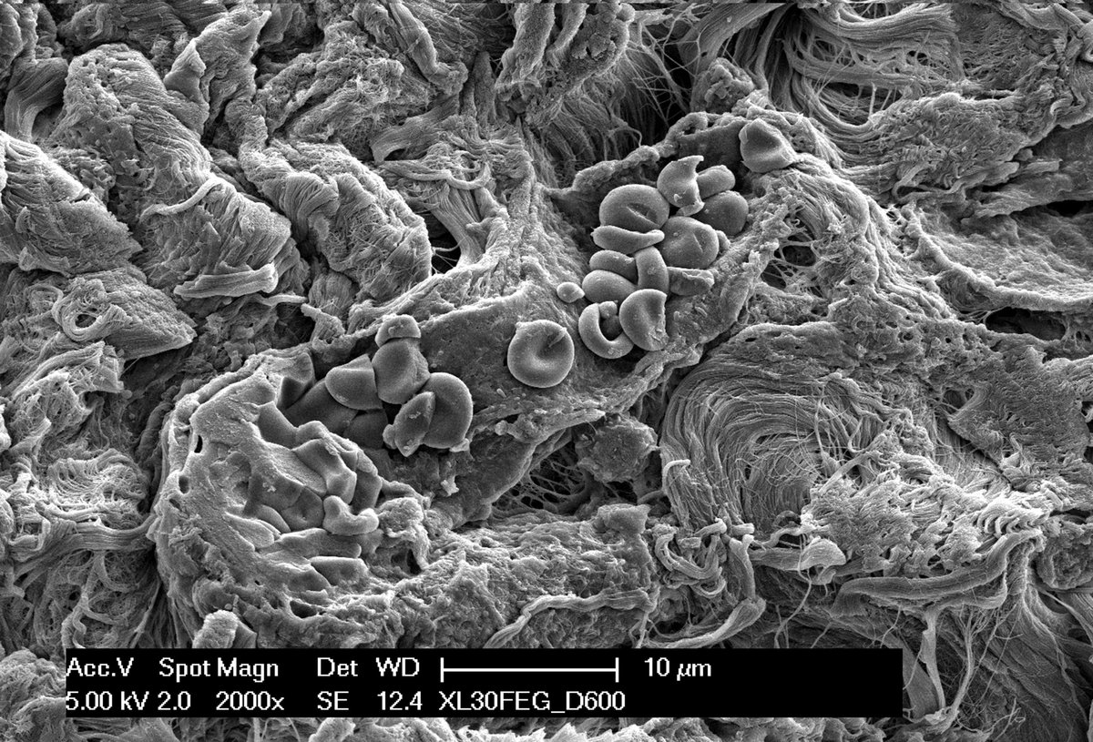
N. 23 - Erythrocytes Colorado Boat!
Image acquired at the scanning electron microscope of a newly formed vessel full of erythrocytes inside a collagen scaffold loaded with Dental Pulp Stem Cell Hypoxic Secretome. Scale bar 10 µm
-
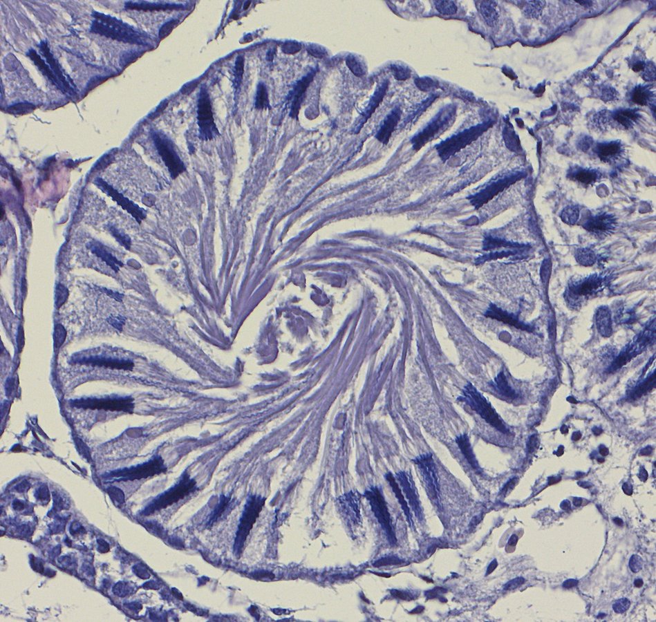
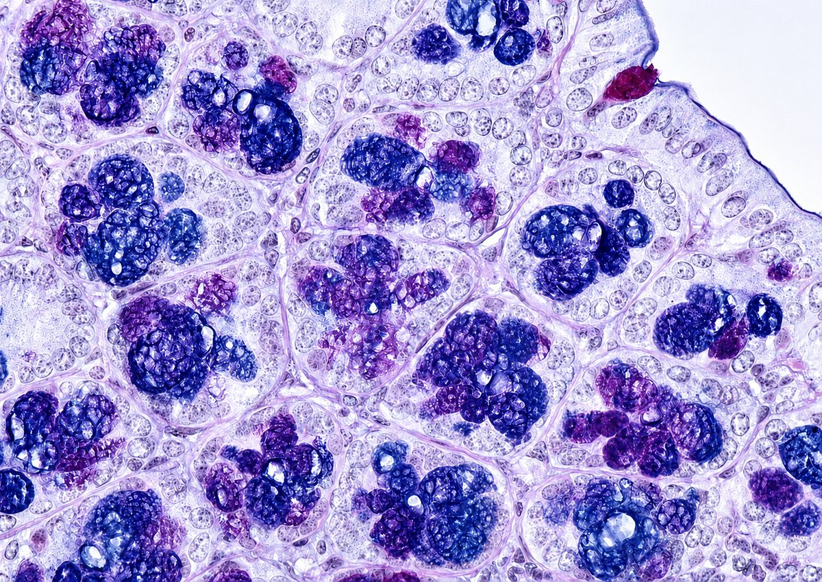
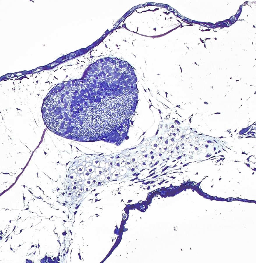
N. 24 - The geometry of reproduction (la geometria della riproduzione)
Cisti spermatica di Mustelus mustelus (squalo palombo) caratterizzata da spermatozoi maturi con teste disposte radialmente alla periferia della cisti e flagelli organizzati in una struttura spiraliforme centrale